Fig. 5. Ribbon diagram showing the tertiary structure of the cGnIHR2 protein in front (A), top (B) and bottom view (C) with N and C terminals.
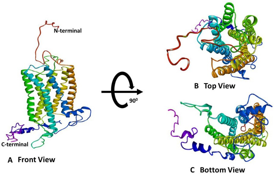
Fig. 5. Ribbon diagram showing the tertiary structure of the cGnIHR2 protein in front (A), top (B) and bottom view (C) with N and C terminals.